
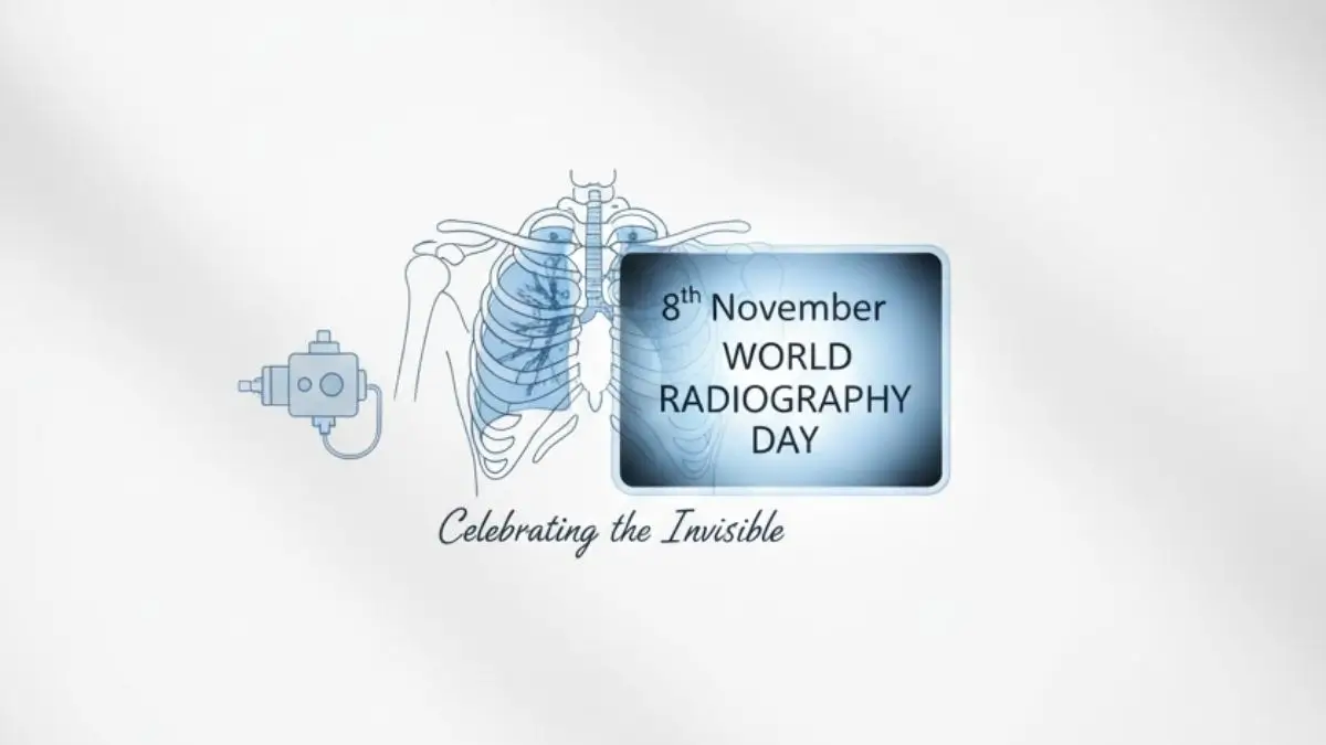
World Radiography Day 2025, observed on November 8, celebrates the revolutionary discovery of X-rays and honors the contributions of radiographers and radiologists to modern medicine. The day highlights how medical imaging—from X-rays to AI-driven diagnostics—has transformed healthcare by enabling early detection, accurate diagnosis, and advanced treatment planning for millions of patients worldwide. This global observance not only commemorates Wilhelm Conrad Roentgen’s discovery in 1895 but also showcases how technological innovation continues to enhance patient safety, clinical efficiency, and the overall quality of healthcare delivery.
History: The Discovery that Changed Medicine
The origins of World Radiography Day trace back to November 8, 1895, when German physicist Professor Wilhelm Conrad Roentgen discovered X-rays at the University of Würzburg. While experimenting with cathode rays, Roentgen noticed an invisible form of radiation that could pass through solid objects and project internal images onto photographic plates.
Key Milestones
- 1895: Roentgen discovers X-rays, transforming science and medicine.
- 1896: Surgeons begin using X-rays to locate fractures and foreign bodies.
- World War I: X-ray units aid battlefield medicine, diagnosing injuries quickly.
- 2007: The International Society of Radiographers and Radiological Technologists (ISRRT) officially designates November 8 as World Radiography Day, aligning with Roentgen’s discovery anniversary.
This breakthrough became the foundation of modern diagnostic imaging, earning Roentgen the first Nobel Prize in Physics in 1901.
Radiographers’ Vital Role in Modern Healthcare
Radiographers are the frontline professionals of diagnostic imaging. They operate sophisticated equipment, ensure radiation safety, and produce high-quality images that guide physicians in diagnosing and treating diseases.
Core Responsibilities
- Operating Imaging Systems: X-ray, CT, MRI, PET, SPECT, and Ultrasound.
- Ensuring Patient Safety: Correct positioning, dose minimization, and radiation protection.
- Supporting Diagnosis: Producing clear, accurate images for fractures, tumors, infections, and organ disorders.
- Cancer Treatment: Collaborating with oncologists in radiation therapy, delivering precise doses to destroy malignant cells while protecting healthy tissue.
- Ethical & Empathetic Care: Balancing technology with patient comfort and compassion.
- Radiographers are indispensable to clinical decision-making, bridging the gap between technology and patient care.
Advancements in Imaging Technology
Since Roentgen’s discovery, imaging has evolved far beyond static X-rays. Today’s radiology combines physics, computing, and artificial intelligence to deliver faster, safer, and more informative images.
Modern Innovations
- Hybrid Imaging: Integration of PET/CT, PET/MRI, and SPECT/CT enables visualization of both anatomical structures and physiological activity in a single scan.
- Functional Imaging: Detects metabolic and molecular changes, improving early disease detection.
- AI in Radiology: Artificial intelligence now assists in analyzing thousands of images within seconds, identifying patterns that may escape human observation.
- Explainable AI (XAI): Adds transparency to diagnostic algorithms, allowing clinicians to understand and trust machine-generated results.
These innovations are transforming radiology into a data-driven specialty, enhancing diagnostic accuracy, reducing human error, and enabling personalized medicine.
Health Impact: Balancing Benefits and Safety
Radiology plays a crucial role in early detection, disease management, and therapeutic planning. Techniques such as X-ray, CT, MRI, ultrasound, and PET scans have become cornerstones of modern diagnostics. However, as the use of ionising radiation increases, safety considerations remain vital.
Health & Safety Points
- Controlled Exposure: Diagnostic imaging uses minimal, regulated doses of radiation.
- Risk Factors: Repeated or unnecessary scans can slightly increase the risk of long-term effects such as radiation-induced cancers or genetic mutations.
- Vulnerable Groups: Children, pregnant women, and patients undergoing multiple scans require extra caution.
- Protective Measures: Use of shielding, optimized protocols, and ALARA (As Low As Reasonably Achievable) principles minimize risks.
With strict adherence to safety protocols, the benefits of radiological imaging far outweigh its potential risks.
Quick Facts
- Date Observed: November 8, annually
- Purpose: Celebrate X-rays and honor radiographers/radiologists
- First Discovery of X-rays: November 8, 1895, by Wilhelm Conrad Roentgen
- First Nobel Prize for Physics (Roentgen): 1901

 CISF Raising Day 2026: Date, History &am...
 International Women’s Day 2026: Date, Th...
 Women’s Day Special: Recent Awards and H...