
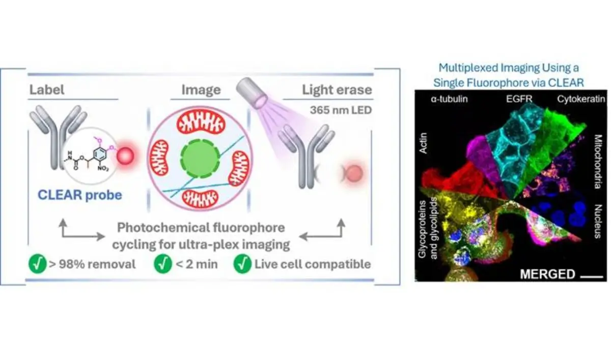
A scientific breakthrough comes as Indian researchers have developed the advanced imaging platform which called CLEAR (Cleavable Light-Erased Antibody Reporter). This will significantly improve the diagnosis of the diseases such as cancer, neurological disorders and immune related conditions. It is developed by scientists at Jawaharlal Nehru Centre for Advanced Scientific Research (JNCASR), Bengaluru and it will allows the researchers to visualize the unprecedented number of proteins within the same biological sample using just a single fluorescent marker.
What is CLEAR Technology?
CLEAR (Cleavable Light-Erased Antibody Reporter) is the novel imaging technology which is designed to map the proteins inside biological samples with exceptional detail.
Proteins are among the most important molecules in the human body because they control nearly every biological function including the cell communication, immune responses, growth and repair.
In disease diagnosis, specially in cancer and neurological disorders, understanding where the proteins are located and how they interact inside the tissues is critically important.
Traditional this imaging methods struggle to visualize large numbers of proteins simultaneously and CLEAR solve this challenge.
Why Protein Mapping Matters in Disease Detection
Proteins serve as the key disease indicators.
A detailed protein map can help the doctors and researchers understand,
- How the cancer cells behave
- How immune cells respond to any infections
- What changes occur in to neurodegenerative diseases
- Which therapeutic targets may work best for individual
This field is called the spatial proteomics, where scientists study the protein identity and exact location inside tissues.
Accurate protein mapping can significantly improve the early diagnosis and treatment decisions.
How CLEAR Technology Works
The CLEAR platform uses the clever ‘erase and reuse’ concept. Think like as a chalkboard.
Scientists first label the specific proteins inside a biological sample by using fluorescent tags.
The sample is then imaged to be under a microscope.
After imaging, a gentle 365 nm LED light pulse is used to erase the fluorescent signal without damaging any part of the sample.
This same sample can then be relabelled with the new protein target and imaged again.
This cycle can be repeated multiple times.
This allows the scientists to create extremely detailed protein maps from the same sample.
What Makes CLEAR Different from Existing Technologies?
Current multiplex imaging methods faces the major limitations.
These includes the,
- Requirement of a multiple fluorophores
- Limited protein detection capacity
- High technical complexity
- Slower imaging workflows
- Difficulty with the fragile biological samples
CLEAR offers the major advantages.
Key Benefits Are,
- Uses only one fluorophore
- Supports the high multiplex imaging
- Works with a live cells
- Provides high spatial resolution
- Faster imaging workflow
- Compatible with a delicate biological tissues
- Enables virtually unlimited protein visualization
This makes the CLEAR as a potentially transformative innovation.
Who Developed CLEAR Technology?
The technology was developed by the researchers at Jawaharlal Nehru Centre for Advanced Scientific Research (JNCASR), Bengaluru which is an autonomous institute under the Department of Science and Technology (DST).
The research team was led by the Prof. Sarit S. Agasti.
Key contributions included,
- Probe design and a synthesis
- Imaging workflow development
- Experimental validation
- Data analysis
Researchers from the Indian Institute of Science (IISc), Bengaluru has been collaborated in testing the platform in complex biological systems and specially immune cell applications.
The findings were published in the prestigious journal Chemical Science, which is published by the Royal Society of Chemistry.
Potential Applications in Healthcare
CLEAR could significantly improve the biomedical research and diagnostics.
Major Application Areas,
- Cancer Diagnosis
- Neurological Disorders
- Immunology
- Drug Development
- Precision Medicine
What is Spatial Proteomics?
Spatial proteomics is an advanced field which studies the proteins in their exact biological location.
Unlike the standard protein analysis, which only identifies a protein presence, spatial proteomics reveals the,
- Where proteins are located
- How they interact
- How disease alters tissue organization
This field is becoming central to the next generation diagnostics.
CLEAR directly supports this growing global scientific area.








 ISRO Successfully Completes Crew Moduleâ...
 Elon Musk Shows Tesla Roadster Performin...
 TRISHNA Satellite Mission 2027: ISRO-Fra...